
A combination of the state-of-the-art techniques to infer ear structure and materialsWe are unravelling the anatomy and physiology of the bush-cricket ear using a range of techniques. Alongside our established micro-CT facilities, we are now including histological approaches of fixed tissues to determine the chemical composition and arrangement of individual membranes within the ear. These will allow us to produce 3D confocal microscope images to complement those from our micro-CT. We are also using molecular biology to identify a range of mechanosensitive proteins within the bush-cricket ear, which will enable us to localise these proteins to specific sensory cells. Finally, we have established a working relationship with the Diamond Light Source national synchrotron facility, which we will continue to visit for nano-CT and radiology driven experimentation.
|
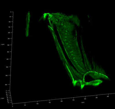
3D view of the crista acustica of Phugis poecila (a transparent bush-cricket) using confocal microscopy
|
Atomic Force Microscopy
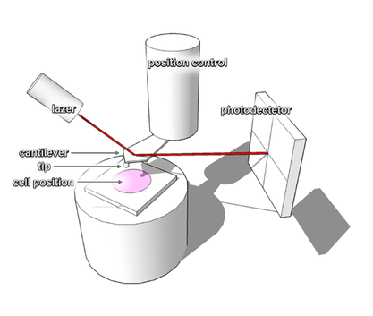 Schematic of AFM instrumentation. With the aid of an inverted microscope lased alignment and sample positioning are achieved. The sample is maintained in near physiological environment by performing experiments in a fluid chamber.
We are investigating the mechanical properties of the acoustic components of insects using Atomic Force Microscopy (AFM)-based Nanoindentation. By using the AFM as a force spectroscopy tool, a series of force versus displacement curves is generated that can be used for further analysis using appropriate mathematical models to calculate the elasticity. Mechanical characterisation of the materials associated in sound transmission and processing facilitates the development of new 2 & 3D models simulating the auditory processes. For example, the insect trachea is a hollow microscale tube that is normally subjected to complex mechanical loading, while its mechanical properties and inner architecture will influence the processing of sounds travelling along its length. In addition, with the aid of AFM we are aiming to associate the frequency analysis of single cells in the crista Acustica with their mechanical properties.
Our primary goal is to quantify the elastic properties of the sound transmission pathways of the tracheal tubes and the mechanics of the transduction organs mainly comprised of the tympana membranes and inner ear cochlea. We are using a variety of AFM cantilevers to achieve the force resolution required for mechanical characterization of the various hearing components, which can be from trachea tissues down to single cochlear cells. By using the spring stiffness of the cantilever we can extract information about the elastic properties of the samples.
In addition, we are using a variety of cantilever tips to indent the materials under consideration. Although AFM based nanoindentation is generally conducted using sharp tips, we are also using in-house modified cantilevers with spherical microspheres that are more appropriate to test soft single cells. Position precision is achieved by using a fine piezo motor that moves and retracts the base of the cantilever towards the surface of the sample in vertical direction, while the laser deflection on top of the cantilever is measured continuously by a photodetector.
In addition, we are using a variety of cantilever tips to indent the materials under consideration. Although AFM based nanoindentation is generally conducted using sharp tips, we are also using in-house modified cantilevers with spherical microspheres that are more appropriate to test soft single cells. Position precision is achieved by using a fine piezo motor that moves and retracts the base of the cantilever towards the surface of the sample in vertical direction, while the laser deflection on top of the cantilever is measured continuously by a photodetector.

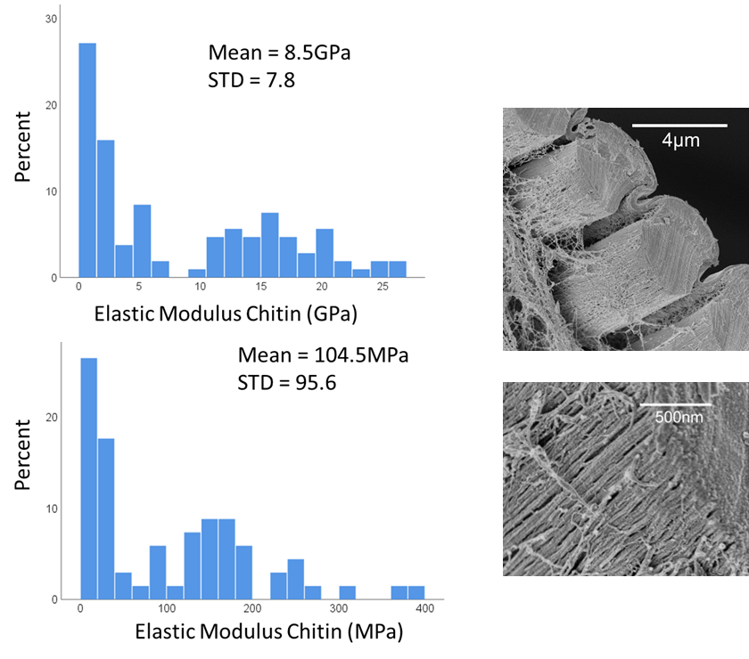
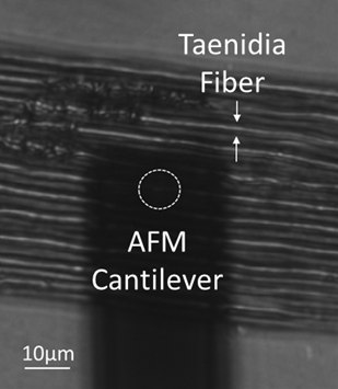
Nanomechanical analysis revealed the complex intrinsic organization of the taenidia, associated with an intricate interplay between chitinous nano-fibrils and resilin proteins. This work highlighted the key role of resilin in the evolutionary optimisation of the fibre-composite tube and the functional reinforcement-recovery adaptation. Overall, we have managed to elucidate the nanoscale mechanics of composite biomaterials with an aim to develop biomedical and bio-inspired applications [Siamantouras et al 2022, ActaBiomaterialia, open access article here).
High Resolution Microscopy of Auditory Structures
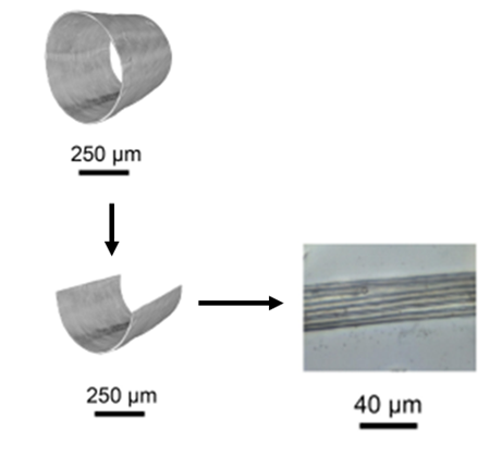
Sound waves are transmitted to the tympanic membrane via the trachea, a hollow tube that is located in the legs of the insect. Due to this position, trachea was developed to sustain loading and is distinguished by an intricate interior architecture and complicated mechanical behaviour. To clarify the organisational structure of the trachea we are using various high resolution microscopy methods, such as Electron Microscopy and Synchrotron X-ray imaging.
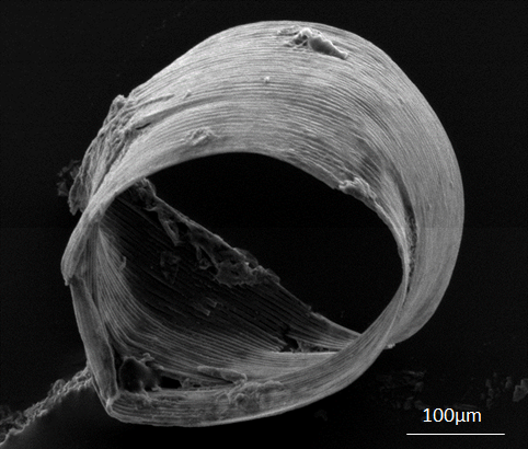
Electron microscopy of Gryllus bimaculatus trachea showing the structure and shape of the dissected hollow tube.
|
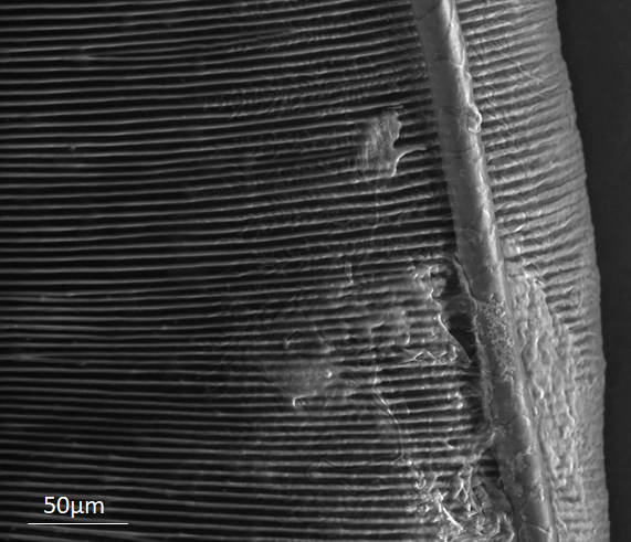
Electron microscopy of a Gryllus bimaculatus trachea showing the organization of taenidia fibers and a smaller respiratory tube.
|
|
This project has received funding from the European Research Council (ERC) under the European Union’s Horizon 2020 research and innovation programme.(Grant agreement ERC CoG 2017 773067) |